")
")
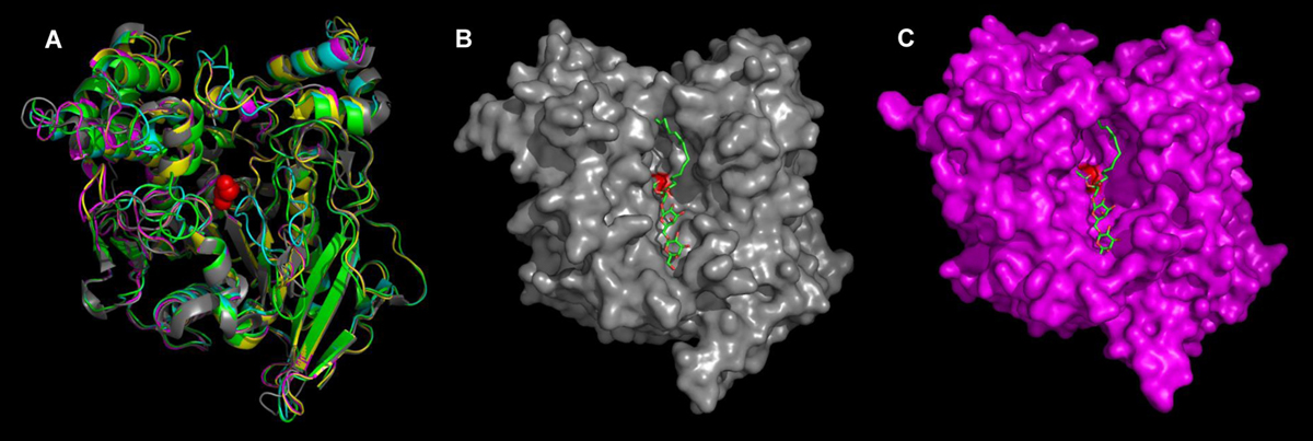
Fig. 8
Download original image
Three-dimensional molecular models of pancreatic carboxylesterases from Mugil cephalus. (A) 3D models, presented in "ribbon" form, of McCEH1 (green), McCEH2 (yellow), McCEH3 (magenta), and McCEH4 (cyan), superimposed onto the known 3D structure of human pancreatic HCEH (grey; (Moore et al., 2001)). The atoms of the catalytic serine are shown as red spheres. (B) Structure of HCEH showing molecular surfaces (grey) and a DGDG molecule (green stick model with oxygen atoms in red) positioned in the active site by molecular docking. (C) 3D model of McCEH3 showing molecular surfaces (magenta) and the same DGDG molecule, which also fits well into the active site of McCEH3. These images were generated using the PyMol program (Schrodinger, 2010).
Current usage metrics show cumulative count of Article Views (full-text article views including HTML views, PDF and ePub downloads, according to the available data) and Abstracts Views on Vision4Press platform.
Data correspond to usage on the plateform after 2015. The current usage metrics is available 48-96 hours after online publication and is updated daily on week days.
Initial download of the metrics may take a while.