")
")
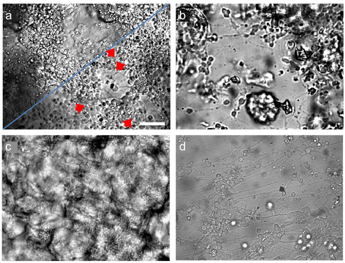
Fig. 1
Download original image
Gel-embedded, gel-invading and gel-covered cortical neurons at 4 days in vitro (DIV). (a) Filtered guar gum gel (< 3%) containing laminin with well differentiated 2D cell carpet at the bottom of the culture well (top left) and embedded neural clusters with a few clearly visible axonal connections in between (red arrows) about 100 μm above the bottom (bottom right). (b) Unfiltered locust bean gum gel (3%) containing laminin with low density network of differentiated neurons at the bottom and cell clusters as well as plant fragments in focus. (c) Center of a cloudily structured gellan gum gel (3%) layer without any added adhesion factors shows less densely, but clearly differentiated neurons interconnecting with less differentiated clusters. Neurons, originally seeded on top of the gel, had sunk or migrated into and homogeneously distributed within the gel. (d) Neurons covered directly after seeding by a sodium alginate gel (4%) with intercalated laminin have differentiated comparable to control cultures (not shown). In all images, undifferentiated cells appear as bright round spheres. Scale bar: 50 μm (a, b, d) and 100 μm (c).
Current usage metrics show cumulative count of Article Views (full-text article views including HTML views, PDF and ePub downloads, according to the available data) and Abstracts Views on Vision4Press platform.
Data correspond to usage on the plateform after 2015. The current usage metrics is available 48-96 hours after online publication and is updated daily on week days.
Initial download of the metrics may take a while.